Necrosis is an irreversible process of destruction and death of cells, human organs, which is caused by exposure to pathogenic bacteria. The cause of development may be: exposure to high temperatures (with a burn), chemical or infectious agents, mechanical damage. Necrosis can be coagulation (dry) or collication (wet). The article will examine in more detail the causes of dry necrosis, as well as methods for its treatment.
What is coagulation necrosis
Dry necrosis often affects organs rich in protein, but with a low liquid content. These include:
- kidneys
- adrenal glands;
- spleen;
- myocardium.
The death of organ cells occurs due to insufficient blood supply and oxygen enrichment as a result of thermal, chemical, mechanical, toxic damage. As a result, dead cells dry out and a mummification process takes place. Dead cells are distinguished from living cells by a clear line.
Causes of dry necrosis
Dry necrosis is formed if:
- there was a process of violation of blood supply to a specific site of a certain organ, as a result, a deficiency of oxygen and necessary nutrients arose;
- the disease developed gradually;
- the affected parts of the organs did not have enough fluid (fat, muscle tissue);
- pathogenic microbes were absent in the cell lesion zone.
The development of dry necrosis is more prone to people with strong immunity and malnutrition.
Coagulation necrosis: a developmental mechanism
Due to insufficient oxygen saturation of cells and circulatory disturbances, the process of coagulation and compaction of protoplasm takes place, then the affected area dries up. Damaged parts have a toxic effect on neighboring living tissue.
The affected area has a characteristic appearance: dead cells are outlined by a clear line and have a pronounced yellow-gray or clay-yellow color. This area is compacted over time. When cut, you can see that the fabrics are absolutely dry, have a curdled consistency, while the pattern is fuzzy. As a result of the decay of the nucleus, the cells look like a mass of homogeneous cytoplasm. Further, with the development of necrosis and inflammation, one can notice the rejection of dead tissue. If the disease affects the auricle or bones of a person, a fistula is formed. However, the mechanism of development of coagulation necrosis is not yet fully understood.
Varieties of coagulation necrosis
Coagulation necrosis includes several types:
- Heart attack is the most common form. It develops due to coronary disease. Does not develop in brain tissue. With a heart attack, complete regeneration of damaged tissues is possible.
- Waxy (Tsenkerovsky) - develops as a result of severe infectious damage. The disease affects muscle tissue, often leading to the muscles of the thigh and the anterior abdominal wall. The development of necrosis is provoked by previous diseases, such as rash or typhoid fever. Affected areas are gray.
- Caseous necrosis is a specific type of disease. The companion of tuberculosis, syphilis, leprosy, leprosy, Wegener's disease . With this type of necrosis, death of the stroma and parenchyma (fibers and cells) occurs. The peculiarity of this disease is that, in addition to dry areas, pasty or curdled granulomas are formed. The affected tissue has a bright pink color. Caseous necrosis is one of the most dangerous species due to the fact that it is able to "kill" huge areas.
- Fibrinoid - a disease in which the connective tissue is damaged. Necrosis develops with autoimmune diseases, such as lupus or rheumatism. The disease most severely affects the smooth muscles and fibers of blood vessels. Fibrinoid necrosis is characterized by a change in the normal state of collagen fibers and the accumulation of necrotic material. Microscopic examination of the affected tissue is similar to fibrin. In this case, the dead have a bright pink color. Areas affected by fibrinoid necrosis contain a large amount of immunoglobulin, as well as the breakdown products of fibrin and collagen.
- Fat - the disease is formed as a result of bruises and hemorrhages, as well as with destruction in the tissues of the thyroid gland. With necrosis, the peritoneum and mammary glands are affected.
- Gangrenous - can be dry, wet, gas. Pressure ulcers in bedridden patients also belong to this type of necrosis. Most often, bacteria that enter the affected areas contribute to the onset of the disease.
Dry gangrene as a form of coagulation necrosis
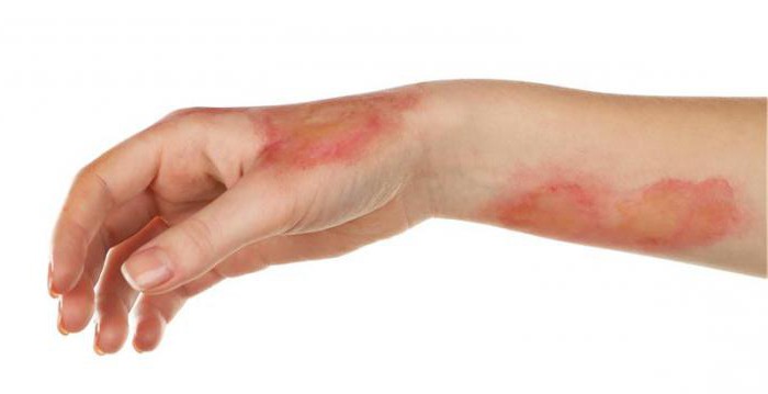
Dry gangrene is a disease in which necrosis of the skin in contact with the external environment develops. As a rule, any microorganisms are not involved in the development of the disease. Dry gangrene most often affects the limbs. Damaged tissue has a dark, almost black color and a brightly contoured outline. Color changes under the influence of hydrogen sulfide. This happens because hemoglobin pigments are converted to iron sulfide. Dry gangrene develops under the following conditions:
- With arterial thrombosis and atherosclerosis of the limbs.
- When exposed to limbs of high or low temperatures (with burns or frostbite).
- With the development of Raynaud's disease.
- In the presence of infections, such as typhus.
Treatment is carried out only by surgical removal of dead tissue.
Wet gangrene
Wet gangrene is a disease that develops when a bacterial infection gets on damaged tissue. The disease affects organs rich in moisture, can occur on the skin, but more often spreads to internal organs. Wet gangrene affects the intestines (with obstruction of the arteries) and the lungs (occurs as a consequence of pneumonia).
Often, the disease occurs in children, since their immunity when an infection is attached is more susceptible to the formation of gangrene. The soft tissues of the cheeks and perineum are affected. This disease is called water cancer. Affected areas become very swollen and have a dark color. There is no boundary line, so the disease is difficult to surgical treatment, since it is difficult to determine where the affected tissue ends. Gangrenous areas have a very unpleasant odor, and the disease often leads to death.
Gas gangrene and bedsores
Gas gangrene in its manifestations is very similar to wet, but the reasons for the development are different. This type of gangrene develops if bacteria of the species Clostridium perfringens get on the tissues affected by the beginning necrosis and actively multiply. Bacteria in the process of their life emit a specific gas, which is found in the affected tissues. Mortality in this disease is very high.
Pressure ulcers belong to one of the types of gangrene, in which the process of tissue death occurs. Diseases are most prone to bedridden patients, as certain parts of the body are under pressure due to prolonged immobilization and do not receive the necessary substances together with blood. As a result, skin cells die. The most affected area of the sacrum, heel, femur.
Diagnosis of coagulation necrosis
To make a diagnosis of “coagulation necrosis”, if the lesions are superficial, it is enough for the doctor to take blood and a sample of the damaged tissue for analysis.
If there is a suspicion of organ necrosis, the examination is carried out more extensive. To do this, you must:
- Take an x-ray. This study is especially relevant if there is a suspicion of gas gangrene.
- Carry out a radioisotope study. It is prescribed if the x-ray has not revealed any changes (at the initial stage of the disease). A radioactive substance is introduced into the human body. If there is a necrotic change in the tissues of the organ, then it will be highlighted with a dark spot.
- Have a CT scan. It is carried out if there is a suspicion of bone damage.
- Perform an MRI scan. The most effective research method, since it shows even minor changes associated with impaired blood circulation.
Complications of Necrosis
Necrosis is the "death" of damaged organs and tissues. Therefore, its various types, such as heart attack, necrosis of the brain, kidneys or liver, can lead to death of a person.
Extensive necrosis can also lead to serious complications, for example, with multiple bedsores, a dangerous infection can be attached. Dead tissues secrete the products of their decay into the body, thus leading to toxic complications. Even milder forms of the disease can lead to unpleasant consequences, for example, the formation of scars in the myocardium or the formation of cysts in the brain.
Necrosis treatment
Treatment of necrosis begins with determining its type, assessing the damage it causes, and identifying concomitant diseases.
When diagnosed with dry skin necrosis, local treatment is prescribed:
- Treatment of affected areas with brilliant green.
- Cleansing the skin with antiseptics.
- Bandage with a solution of "Chlorhexidine."
The patient is prescribed medical and surgical treatment in order to restore normal blood circulation, including in the affected areas. To remove dead cells, a surgical operation is most often performed to resect the affected areas. Amputation of the extremities is carried out in order to protect healthy areas from the spread of the disease.
Dry necrosis of internal organs is treated with anti-inflammatory drugs, vasodilators, chondroprotectors. In case of treatment failure, surgical treatment is performed.