Holt-Oram syndrome is a serious disease with an autosomal type of inheritance, which is characterized by the defeat of several genes at once. They are responsible for the anatomical structure of the heart and skeleton. This ailment is called hand-heart syndrome.
What it is?
This inherited disease combines varying malformations of the upper extremities and congenital heart defects. It is very important to recognize the disease at the beginning of its development. To do this, carry out genetic research. They not only confirm the diagnosis, but also predict the likelihood of the disease in future generations.
Ultrasound examination during pregnancy makes it possible to timely detect violations in the development of the skeleton. If indications match the signs of the disease, an intrauterine scan of the heart of the unborn child is performed. Usually, Holt-Oram syndrome is determined after the birth of the baby. Suspicious defects in the structure of the skeleton are carefully examined by a cardiologist, since the severity of the disease depends on the cardiac manifestations of the syndrome.
Birth defects of the disease
Anomalies in the structure of the forearm and hand are characteristic, which appear on one side or both at once. The degree of underdevelopment and deformation of the limbs is different: this is the presence of three phalanges on one finger or when the thumbs are completely absent. Most often this happens with fingers on the left hand. Severe cases occur when there is a radial bone hypoplasia or its complete absence. Much less often note: fingers joined together, deformation of the sternum, underdevelopment of the clavicles and shoulder blades, curvature of the spine. Skeletal changes do not pose a threat to human life. But a violation of the functional ability of the affected organ complicates life.

About 85% of patients suffer from congenital heart defects: narrowing and open duct of the aorta, stenosis of the pulmonary artery, impaired valve structure. Often there is a congenital atrial septal defect. It varies in severity: from barely distinguishable to an extensive secondary defect, the consequence of which is heart failure.
Autosomal dominant type of inheritance
This disease is characterized by:
- Regular transmission of the disease from one generation to another.
- If the gene is highly manifest, the disease does not skip a generation, but can be traced to family members in each genus.
- The gene is passed from parents to children. An exception is the first occurrence of a disease in the family due to a spontaneous mutation of a gene, which is subsequently also transmitted from a sick ancestor to a healthy offspring.
- More often, the disease is transmitted along one parent line.
- Not everyone in the family inherits the disease gene: in healthy family members, offspring are born without signs of manifestation of the disease.
- If the children have one mother and the fathers are different, or vice versa, the father is one and the mothers are different, the children of a common parent can inherit the disease.
- The autosomal dominant type of inheritance does not depend on a person's belonging to a particular gender. Men and women are affected equally by the disease.
Interatrial septum
If a defect is observed, a congenital heart disease develops, which can be isolated or combined with other diseases inside the heart. An atrial septal defect develops when an open message is formed between two atria: left and right. If a patient has Holt-Oram syndrome, he has shortness of breath, heart murmur, fatigue and physical development lag increases, skin pallor and frequent respiratory diseases are observed. Congenital heart disease due to this pathology is observed in approximately 5-15% of patients, moreover, in women it is twice as common as in men.
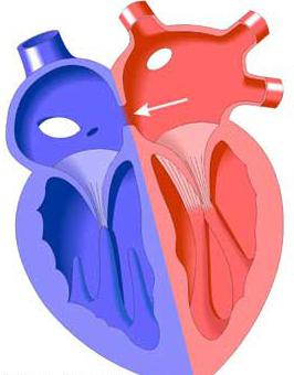
The fact is that the fetus, being in the womb, has an open oval- shaped opening in the interatrial septum. It provides normal blood circulation. After the baby is born, the opening in most newborns closes. Although there are times when this does not happen. But the discharge of blood through this hole is so insignificant that a person does not feel anomalies and does not even suspect that he has it. He lives quietly until old age. Such hereditary anomalies, such as a defect of the interatrial septum, can be of various sizes, shapes and are found in its different departments.
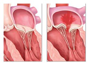
Atrial defects. Types
- An anomaly of the primary part of the septum is a type of abnormal development of the pads and is localized in the lower part of its third next to the tricular valves. They, in turn, can be deformed and not cope with the performance of the intended function. There are cases when with their help a common valve forms with the atrioventricular septum.
- The interatrial septum has a defect in its secondary part, which is observed at the top or in the oval fossa. In this case, blood enters the right atrium from the left. Often these defects are called secondary and they occur when there is a lack of septum tissue. But at the same time, the functional and anatomical patency of the defect is preserved.
Symptoms of Atrial Defects
- Holt-Oram syndrome is directly related to the age of the patient, the size of the septal defect, and the resistance of the pulmonary vessels. Many patients with this defect look completely healthy and do not complain. The fact is that blood can be discharged from left to right due to excessive physical exertion or fatigue, and not due to a defect. In the vast majority of cases, heart murmur is not heard. This sometimes happens with a significant atrial septal defect.
- This anomaly often causes mitral valve prolapse. In patients with such a disease, noise or a click is well audible.
- If the size of the defect is very large, a person develops tachycardia, shortness of breath, cardiac hump, when the boundaries of the organ are extended to the right. You can easily, using your fingers, determine the pulsation of the pulmonary artery.
- A secondary atrial septal defect in rare cases can cause heart failure. This is observed in 3-5% of patients, mainly in children under one year of age. If you do not have surgery, the risk of death is great.
Atrial defects. Causes
Abnormalities occur when the primary or secondary septa and zardocardial ridges are underdeveloped during embryonic development.
- The occurrence of the defect is associated with genetic, physical, environmental and infectious factors.
- Abnormal development of the septum of the child in the womb has a high degree of risk in families whose relatives are sick with congenital heart disease.
- Diseases of the viral nature of a pregnant woman can cause an atrial septal defect. These are rubella, herpes, chickenpox, syphilis.
- Drugs and alcohol during pregnancy.
- Harmful substances in production, radiation exposure.
- Complications due to toxicosis, as a result of which the bearing of the child is compromised.
Atrial septal defects. Classification
In the period from the third to eighth weeks of pregnancy, the fetal heart is laid in the womb. If an incomplete septum forms between the right and left halves of the heart, this leads to the appearance of defects of the atrial septum, which are distinguished by the size, number and location of the holes. The classification of congenital heart defects is as follows:
- Primary defects are characterized by significant sizes: from three to five centimeters. The place of its localization is the lower part of the septum, which is located above the atrioventricular valves, just above them. They have no bottom edge.
- Secondary defects are formed as a result of the underdevelopment of the secondary septum. They are small in size and are located in its central part or in the region of the mouths of vena cava. It happens that defects of the septum and abnormal flow into the right atrium of the pulmonary veins are combined. In this case, the interatrial septum remains in the lower part.
- Cases are observed when secondary and primary defects are present simultaneously.
- The atrial septum may be completely absent. This leads to the formation of a common, single atrium, which is called the three chamber heart.
Congenital heart defect. Diagnostics
If this disease is suspected in a newborn, an urgent call to a cardiologist is necessary. The specialist examines the symptoms of the disease, evaluates the nature of the pulse and pressure, the condition of all organs and systems. After examining the baby, an electrocardiogram, an ultrasound scan, a phonocardiogram, and an x-ray of the heart are performed. In the case of a severe form of the disease, the diagnosis of congenital malformations is supplemented by another examination - cardiac catheterization, when a probe is inserted into its cavity.
Very often, the parents of the baby are outraged that the attending physician did not detect this dangerous disease when the expectant mother spent the entire period of pregnancy with him. The cause of congenital heart disease can be:
- The low level of professionalism of the doctor.
- Congenital diseases are not always able to be diagnosed on time due to the structural features of the heart and blood vessels of the fetus.
- Imperfect medical equipment.
Causes of Congenital Heart Diseases
Congenital heart defects in newborns are the result of anatomical defects of the heart, its valves and blood vessels, which can occur even in the womb. At first, they may be invisible, or they may disturb the baby immediately after birth. Heart defects are the most common congenital malformations in newborns and make up about 30%. They are the most common cause of death. Causes of the development of the atrial septal defect:
- Viral diseases during pregnancy, such as rubella, measles and others.
- Genetic mutation caused by the future mother's use of drugs, alcohol, nicotine.
- Various deviations of the chromosome sets of parents.
- Irradiation by ionizing radiation.
- Deficiency of trace elements and vitamins in the diet of a pregnant woman.
- The effects of drugs on the fetus.
Symptoms of Congenital Heart Disease
Babies born with this disease are different. Their skin, lips, and auricles are bluish or blue. Sometimes, on the contrary, the baby's skin turns pale, and the legs become cold. This is called a white malformation of the newborn. There are cases when, when listening to a specialist, he clearly hears a noise in the heart.
Very often, a congenital organ disease is imperceptible immediately after the baby is born. Within 10 years, the child will look completely healthy. But, then the disease will begin to appear in the form of blanching or blue skin. Deviations in physical development will become noticeable, shortness of breath will appear.