Nail melanoma or subungual melanoma (lat. "Melanoma", from the ancient Greek "μέλας" - "black" + "-ομα" - "tumor") is a malignant disease that develops from specialized skin cells (melanocytes) that produce melanins. It occurs not only on the inside of the hand and the sole of the foot, but also on the nails (most often the nail of the thumb or toe is affected, but damage to other nails and fingers is not excluded).
How often does it occur?
Among all cancers, the incidence of nail melanoma is about 3% in women, and in men - about 4%. Previously, it was always believed that subungual melanoma can occur mainly in older people, but now this malignant tumor has been observed more often in young people.
Compared to other types of cancer, the growth of nail melanoma is much faster, because the body has a very weak or no response to it. Therefore, according to statistics, after malignant lung tumors, this disease takes the second place.
Kinds
There are several types of subungual melanoma:
- developed from the matrix of the nail (the skin located under the root of the nail, responsible for the development of new tissues);
- emerging from under the nail plate (the main part of the nail that protects the soft tissue of the finger);
- evolved from the skin next to the nail plate.
Causes of nail melanoma
Melanoma of the nail affects people of all races, regardless of country of residence and status. In fact, at present, science has not yet fully established the causes of this disease. However, it is still possible to identify factors that affect the conversion of healthy cells to malignant. Risk groups include:
- people who have fair skin, blue eyes, lots of pink freckles, and fair or red hair;
- those who have a history of sunburn (even if they were received in childhood or adolescence);
- people whose family history has repeatedly recorded cases of subungual melanoma are affected by this disease 3-4 times more often;
- people over the age of 50;
- regularly exposed to ultraviolet rays (including those from artificial tanning equipment);
- those suffering from a lack of vitamins, relaxation and poor immunity, as well as those who work with aggressive environments and chemicals, are at risk. Next, find out what melanoma looks like.
Signs of the disease
In most cases, with the progression of the disease, the symptoms of nail melanoma also change. Therefore, it is extremely important not to lose sight of the first signs characterizing the occurrence of a malignant formation, because, as a rule, the early development of the disease is asymptomatic. But at later stages, the following symptoms begin to appear:
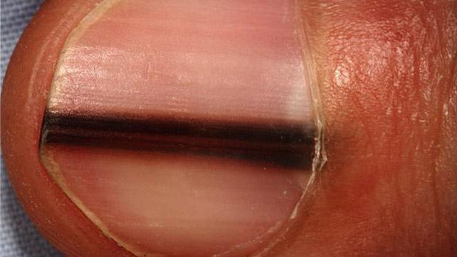
- A dark pigment spot appears under the nail plate. This spot may look like a longitudinal strip on the nail bed. Sometimes the occurrence of nail melanoma may be preceded by a minor injury to the finger of the patient, who did not consult a doctor in a timely manner.
- As a rule, an increase in the spot under the nail occurs within a few weeks or months. It begins to change color to light or dark brown and becomes wider in the area of cuticle growth, and as a result can completely cover the entire area of the nail.
- A malignant neoplasm begins to spread to the nail roll, which surrounds the nail plate.
- Bleeding ulcers and developing nodules begin to appear, which lead to deformations, cracks and thinning of the nail plate. And also from under the nail pus begins to stand out.
So, you already know what melanoma looks like. The above signs will allow the doctor to suspect pathological destruction of epidermal tissues and the presence of this dangerous disease in the patient. In some cases, the specialist examining the patient confuses a dermatological ailment with a panaritium of an infectious nature of origin and prescribes surgical debridement of the affected skin surface.
Precious time is wasted that would have to be used to treat the tumor, and the signs of cancer return again and with a more vivid manifestation of the clinical picture.
Since very often the neoplasms under the nail have no color, in half the cases of this disease, unfortunately, patients notice the external signs too late. Nail melanoma of this kind can be seen only if a nodule begins to form under the plate, which raises the nail up.
It should be noted that both hands and feet are equally affected by this disease. If a malignant tumor has spread to the sole, then it provokes obvious discomfort during movement. In the early stages, this disease is so asymptomatic that sometimes doctors confuse it with ordinary skin warts.
Stages
So, we select all stages of nail melanoma:
- First, damage to the surface of the skin is observed, the nail plate reaches a thickness of 1 mm, however, this does not cause concern to the patient.
- During the second stage, subungual melanoma reaches a thickness of 2 mm and begins to spread along the nail plate, changing the pigmentation. The spot expands, darkening at the same time.
- After this, the malignant cells begin to spread to the nearest lymph nodes, and in this case, damage to the skin around the nail is often observed.
- At the fourth stage, metastases begin to appear in the liver, lungs and bones.
Everyone should remember that it is important to recognize the symptoms of subungual melanoma on time.
Pathology diagnostics
The reason for a visit to a specialist should be any pigmented change in the nail plate, especially if it has increased in size (up to 3 mm or more), because nail melanoma at the early stage often has ambiguous signs. To determine the malignancy of the neoplasm under the nail, qualified specialists use a dermatoscope - a special optical microscope used to scan the stratum corneum of the nail and skin to assess pathological changes visually: the degree of spread, size and thickness of the tumor. Next, you will learn how to distinguish subungual melanoma from hematoma.
Biopsy
If a malignant origin of the tumor was detected during dermatoscopy, then the next step, the doctor prescribes an additional biopsy, which allows you to remove the suspicious formation along with the area of the skin surrounding it and examine in the laboratory a tissue section under a more powerful microscope and determine unambiguously whether it is a malignant tumor or an ordinary hematoma .
Sometimes it happens that a histological examination refutes the presence of nail melanoma in the patient and diagnoses other diseases: the subungual hematoma, which is formed, as a rule, due to bleeding or bruising, fungal infection, purulent granuloma, paronychia, squamous cell carcinoma. If a histological examination still found a malignant tumor, then the final step is an ultrasound examination (ultrasound) of the organs and tomography to exclude the presence of metastases. How long does subungual melanoma develop? About it below.
Nail melanoma treatment
Melanoma, along with part of healthy tissues, as well as subcutaneous fat and muscle, is completely removed (excised) surgically. Sometimes it happens that melanoma has already spread very much. Then, together with it, completely remove the nail plate, and in especially advanced cases, the entire phalanx of the finger or toe is amputated. Also, if the patient is diagnosed with nail melanoma, then he is prescribed a biopsy of the lymphatic tissues, which will help doctors determine the degree of spread of the malignant tumor to the local lymph nodes. Subungual thumb melanoma is common.
If metastases are detected as a result of histological examination, then their removal is additionally carried out. And in addition, complete removal of the lymph nodes is prescribed, and then, depending on the individual characteristics of the patient's body, a complex or combined treatment is prescribed.
Additional methods
Additional methods of dealing with this ailment are:
- Chemotherapy.
- Radiation therapy.
- Laser Therapy
If, apart from the nail plate, nothing was removed, then after the operation to remove the melanoma the nail grows back.
Forecast
If a patient in a medical institution was provided with timely and competent help, then the prognosis for him will be very favorable.
If the patient did not bother to turn to a qualified specialist in time, a visit to which was delayed for a long period, then the tumor can already give metastases and the treatment process in this case is greatly complicated, because the chances of survival are reduced. Approximately 15 to 87% of patients survive for five years after diagnosis.
Therefore, value your health, do not neglect it and immediately consult a doctor with the first symptoms.