This pathology is widely known due to its occurrence in the central nervous system, but it can form anywhere in the body. The genetic predisposition to AVM and the facts of its transmission by inheritance are unknown. It is believed that this is not a hereditary disease.
Arteriovenous malformation is a vascular pathology - abnormal development of the vessels of the nervous system. More precisely, we are talking about a congenital anomaly in the vascular structure of the brain or spinal cord. The term "malformation" in Latin means "poor formation", that is, any deviation from healthy physical development with a gross change in the structure and functions of an organ or tissue is implied. Such a defect may be congenital or acquired as a result of an injury or illness. A similar deviation develops on the skin, in the lungs, and kidneys, but most often it occurs in the nervous system.
What is the essence of the disease?
This deviation is more common in the brain, in the posterior regions of the hemispheres, and the thoracic or cervical spine is often affected. An abnormality is also found in the spinal cord, but this is rare. Arteriovenous malformation varies from one centimeter to a very large size in the cranial cavity.
The resulting defect usually looks like a ball of thin, thinned and convoluted vessels, in which arteries with veins meet directly, which occurs without the participation of capillaries. Thus, arterial blood does not enrich tissues and organs in any way.
This disease is chronic in nature, and it occurs mainly in men at a young age. In one family, such a pathology sometimes manifests itself at once in several members, but it is not considered hereditary. For the first time, pathology appears at the age of ten to thirty years, and its peak falls on twenty years. Violation of vascular formation occurs in the second month of fetal development, and the exact causes of such an anomaly have not been established. The frequency of the disease in the world is one person per hundred thousand people.
Development mechanism
Normally, blood enriched with oxygen flows from the heart to various tissues and organs. At first, it passes through the arteries. Then the artery passes into the arteriole, through which blood already enters the capillaries. Here is the capillary bed in which cell metabolism occurs. Cells take oxygen from the arteries, giving off waste products with carbon dioxide. Then the blood flows through the veins further, from there rising again to the heart.
In the presence of arteriovenous malformation, blood from arteries enters the veins through a tube called a fistula, and hypoxia develops directly in the tissues against the background of this. In the veins through which the blood passes, there is increased pressure.
With the passage of time, fistulas can gradually expand and increase, and arterial walls thicken. If malformation is very developed, then the blood flow in it is strong, and at the same time, cardiac output also increases. In such cases, arteries and veins look like giant pulsating vessels. They are not able to withstand such pressure, because they are not adapted for this, therefore they stretch and often burst. A similar state of blood vessels can be observed in any part of the body. In the event that the malformation affected only the veins, then they speak of the presence of venous angioma.
Varieties
By structure, the following varieties are distinguished:
- Malformation, in which there are no veins in the vascular bundle, there are only arteries.
- A fistulous type of arteriovenous malformation can occur in the dura mater.
- Racemous branched malformation occurs in seventy-five percent of cases.
- Cavernous malformation occurs in eleven percent of cases. It contains only small capillaries, and the arteries and veins are completely absent, the pressure is not broken. A subspecies in this case is telangiectasia.
In terms of size, they emit:
- The smallest in size is micromalformation.
- Miserable malformations that are less than one centimeter.
- Small malformations, whose size is from one to two centimeters.
- Up to four centimeters are medium malformations. The risk of a break is quite high.
- Up to six centimeters are large malformations that are very dangerous.
- More than six centimeters are already gigantic, at the same time they are torn less often, but it is difficult to cure them.
Arteriovenous malformation is divided by the nature of drainage and its localization. It can be located in the cerebral cortex, that is, directly on its surface. In this regard, they are also called cortical. Other forms are internal malformations, which are often localized in the brainstem or in the hypothalamus. An arteriovenous fistula may be located inside the dura mater .
Symptomatic manifestation of brain malformation
Arteriovenous malformation of cerebral vessels, it is also called cerebral, is accompanied by the following main symptoms:
- The appearance of cephalgia of varying intensity, not having any characteristics in regularity and duration. Moreover, the pain does not coincide with the localization of malformation, and its intensity is different.
- The presence of seizures. In this case, general or partial convulsions are observed in various parts of the body. Loss of consciousness is not observed.
- The appearance of dizziness and fainting.
- The development of muscle weakness along with paresis of the limbs.
- In the event that the cerebellum is affected, gait is impaired. At the same time, staggering and lack of coordination are observed.
- In the frontal lobes, loss of vision may be noted.
- The appearance of dysarthria.
Symptoms of arteriovenous malformation of the right parietal lobe may not appear for a very long time and sometimes are accidentally detected during examinations.
Neuralgic symptoms
During the growth of malformation, when pressure on the brain begins, neurological symptoms occur. The following manifestations may be present:
- Intracranial pressure increases, persistent pains in the head are pressing or pulsating in nature.
- The appearance of apathy, lethargy, decreased performance.
- Impaired coordination of movements.
- Decreased intelligence.
- The appearance of speech disorders in the form of motor aphasia.
- Failure of innervation of certain areas of the body.
- Shaky gait with sudden falls on his back.
- The appearance of seizures and muscle hypotension.
- Paresis of limbs.
- Vision problems in the form of strabismus or blindness.
Against the background of the gradual development of arteriovenous malformation of cerebral vessels, neurological symptoms can increase sequentially. Upon reaching middle age, the disease becomes stable, and new disorders no longer occur. Women may feel worse and have new symptoms in case of pregnancy. Hemorrhagic stroke in pregnant women is caused by this disease in twenty-three percent of cases.
Arteriovenous spinal cord malformation
In this case, the symptoms will be as follows:
- Problems with the sensitivity of the limbs, for example, may not feel pain or touch.
- The occurrence of intense pain.
- The appearance of sudden progressive paralysis of the lower extremities. Arteriovenous malformation is a very serious disease.
- The appearance of tingling in the limbs.
- Failure of sphincteric activity and urodynamics when it is impossible to control bowel movements or urination.
Most people recover almost completely after the first attack, but there is a risk of recurring symptoms. In the absence of treatment over time, the patient may become helpless and will completely depend on his loved ones.
Symptoms of rupture of blood vessels
Vascular rupture against the background of malformation is possible in every second patient. A significant role in this is played by increased stress, stress and alcohol consumption. Hemorrhage occurs suddenly. Often it is subarachnoid in nature. Symptoms are similar to a stroke. The patient complains of a severe headache, from which he may even lose consciousness. Without visible prerequisites, vomiting occurs, and after cleansing the stomach, no relief appears. There is a fainting state. There is irritation with pain in the eyes, which is caused due to bright light, impaired vision, complete blindness develops, speech disorders are noted.
Convulsive seizures along with hearing loss are not ruled out. Visual disturbances may occur, limb paralysis develops. When hematomas appear, meningeal syndrome is often diagnosed, while the pressure is increased. The next day the temperature rises. With proper treatment, health improves after five days. After hemorrhage in the first year, the risk of its recurrence remains. Moreover, if you do not carry out treatment, then the risk increases three times.
Diagnostics
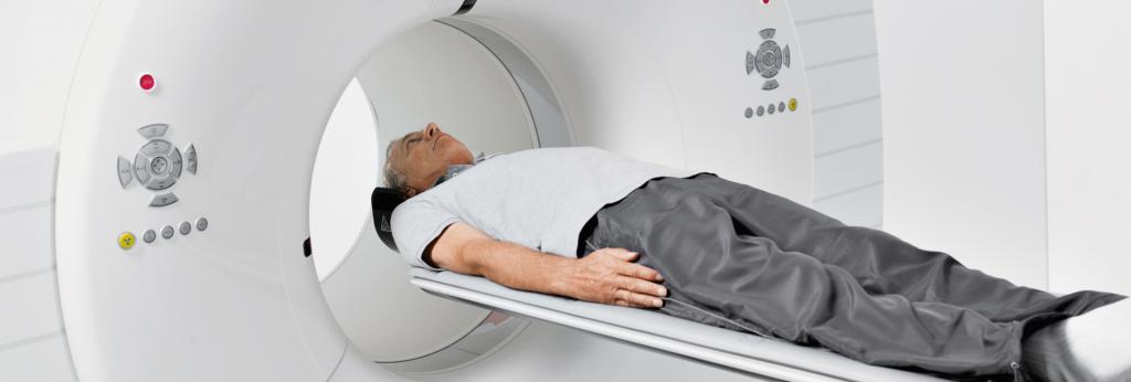
Patients most often seek help after hemorrhage. The doctor conducts a detailed examination of the patient, it reveals the presence of injuries and diseases, determines the neurological status. Then appoint a computer and magnetic resonance imaging and angiogram.
These methods for diagnosing AVMs allow for a layered study of the brain structure, while determining the location of malformation with its size, and assessing the general condition of the brain. With computed tomography, due to x-rays, it is possible to determine the anomaly inside the parenchyma. When performing CT angiography, the arteries of the brain are further detailed. Computed tomography is the fastest method, but not the most effective, it is more suitable for detecting hemorrhage.
MRI with arteriovenous malformation is more informative. Thanks to her, this disease is easily detected and the severity of the patient's condition is determined. A cerebral angiogram is very accurately assessed by the patient’s condition , but this is a rather expensive examination. As part of its conduct, a catheter is inserted into the peripheral artery, which advances to the vessels of the brain. After taking pictures of blood vessels. Although there is a risk of complications after this manipulation, but only this method allows you to accurately determine the causes of hemorrhage.
Electroencephalography determines the foci of excitation, finding the zone of its localization. When performing Doppler ultrasound, doctors determine the speed of blood flow and examine the spatial position of the vessels in the affected area. Angiography is also performed. But this procedure lasts a long time, and they do it under general anesthesia. Angiography is indispensable for determining the venous increase in pressure, it is very important in the choice of surgical treatment of arteriovenous malformation (ICD Q28.2.).
Treatment
There are three options when choosing a treatment method. We are talking about the method of surgery, embolization of arteriovenous malformations and radiosurgical treatment. It is important to determine the degree of risk of the operation and evaluate the possible consequences. The main goal of any technique in this case is to achieve absolute obliteration in order to avoid further possible hemorrhage in the cranial cavity.
Surgical treatment
As part of this method, malformation is completely removed with volumes up to 100 milliliters. Surgical technique involves opening the skull in order to detect malformation. In addition, it is subsequently cauterized by a laser or using other tools. The scorched area is completely removed from the tissues. In the event of a successful operation, the patient recovers completely. But complications, however, are possible in the form of strokes.
After the operation, a full rehabilitation course is carried out for one week. After discharge of the patient, he is recommended treatment with nootropics and angioprotectors. As part of prevention, it is necessary to periodically be examined by a vascular surgeon and a neuropathologist, and magnetic resonance imaging also needs to be done.
Embolization, or Endovascular Surgery
Endovascular surgery involves the removal of malformation from the general bloodstream by gluing vessels. Against this background, complete gluing of blood vessels is feasible in thirty percent of patients, in other patients it is partial. This technique is very often used, and it effectively prevents hemorrhages. The technique of embolization in endovascular surgery involves the supply of a special adhesive element through a catheter.
Radiosurgical treatment
When using this method, obliteration of malformation is possible with its size less than three centimeters. After such an operation, arteriovenous malformation, eighty-five percent of patients recover. This method is used when the localization of malformation is inaccessible for performing the classical operation. As part of the implementation of this technique, radiation is focused that is directed to the place of the anomaly; this procedure lasts exactly one hour. Further, the vessels independently sclerosize for two years and are replaced by connective tissue. The disadvantage of this technique is that before the development of sclerosis, hemorrhages in this area may occur.
Currently, various types of operations are being actively combined. This allows you to expand the possibilities of radical approaches, reducing the percentage of complications.
Arteriovenous spinal cord malformation is treated with surgery. It is also possible to use the intervention method. The latter method is slightly invasive. It involves the introduction of a special adhesive element, which immediately hardens, clogging the vessel. When using this method, there is a risk of damage to healthy vessels. In this regard, the introduction of the substance is carried out as close as possible to the anomaly.
Sometimes special microcoils are introduced, which are adjacent to abnormal vessels and block the access of blood to them. In addition, detachable spirals subsequently assist in the development of collaterals. Blockade of blood vessels is carried out by a substance that resembles particles of sand. But such particles can lead to new recanalization. In this regard, for the purpose of prevention, angiography should be performed annually. In the event that the discovery of malformation occurs, embolization is repeated. It is usually carried out under anesthesia, and the duration of the procedure is from three to six hours. In the event that the patient has a slight soreness after the procedure in the incision area, analgesics are prescribed.
Preventive measures
Arteriovenous vascular malformation is a consequence of abnormal and impaired embryogenesis. In this regard, prevention should be reduced solely to the prevention of gaps that are provoked by a number of factors in the form of heavy physical exertion, stress, smoking, alcohol, high blood pressure, etc. Those people who have already undergone surgery should regularly undergo magnetic resonance imaging.