During the waiting period for the baby, the fairer sex has to undergo numerous studies and pass a lot of tests. So, before each appointment with a gynecologist, the expectant mother must donate blood and urine for examination. The results obtained allow us to judge the course of pregnancy and the state of health of a woman. In this article, we will talk about how long an ultrasound scan is done during pregnancy. You will find out the features of this diagnosis. You can also find out the generally accepted terms for ultrasound during pregnancy.
Ultrasound diagnostics
This type of research has been used for a long time. Every year there is an improvement in inspection methods. So, in modern medical clinics, you can not only go through an ordinary ultrasound scan, but also do a study in several planes (3D and 4D).
Ultrasound diagnostics is carried out as follows. During the study, the doctor applies a special sensor to the patient’s body that sends pulses. This ultrasound is reflected from the organs and gives an image on the screen. The patient and the doctor cannot hear the ultrasound, as it has very high frequencies.
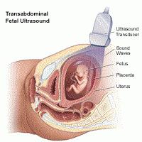
It is worth noting that the study can be carried out in several ways. In the earliest stages of embryo development, a transvaginal probe is selected. The doctor puts on it a special conductive gel and injects into the vagina. Later pregnancy allows for transabdominal ultrasound. In this case, a slightly different sensor is selected, which is applied to the belly of the future mother.
How many times do ultrasound during pregnancy and before it occurs?
If a woman only plans to conceive, then the doctor prescribes her a course of examinations called folliculometry. This method allows you to track the growth of the follicle and determine the exact time the egg leaves it. Thanks to this method, the probability of conception increases several times. During folliculometry, a woman is given one to three ultrasound examinations.
With the onset of pregnancy, the expectant mother should visit the ultrasound room as prescribed by the doctor. How many times do an ultrasound in this case? In the normal course of pregnancy, the study is carried out no more than three or four times over the entire period. However, in half the cases, women are forced to undergo this procedure more often.
Establishment of the fact of pregnancy: for how long?
When can you safely go for an ultrasound to determine the fetal egg in the uterine cavity? What are the terms for ultrasound during pregnancy in this case?
You can establish the completed conception starting from the fifth week of pregnancy. However, you will not be able to see the embryo. However, an experienced specialist will detect a formation in the uterine cavity, which will subsequently become your child. At earlier times, there is a chance of error, as some devices cannot recognize such a small point (during this period, the size of the fetal egg is not more than two millimeters).
What are the terms for ultrasound during pregnancy?
In fact, there is no well-established generally accepted deadline for diagnosis. It all depends on the number of fruits, the course of pregnancy, the health and age of the expectant mother. If everything goes smoothly, then the timing of the diagnosis will be separately set for the first, second and third trimester.
First ultrasound
How long does this study take? Doctors recommend a diagnosis at 11-14 weeks. Such an examination allows you to accurately determine the period. By ultrasound, this is much easier to do than during a gynecological examination. Diagnosis is most often carried out by a transvaginal device, since the uterine cavity is still in the pelvic area. However, if modern equipment is available, a specialist can choose the transabdominal method for research.
A study may also report the number of fruits in the cavity of the genital organ. During this period, the children's place is necessarily inspected, its location and the presence of detachments is noted. At this time, you can identify possible deviations in the development of the embryo. The size of the unborn child is not so small and the main formed organs are quite clearly visible.
Second Ultrasound
2 ultrasound period has the following: from 20 to 22 weeks of embryo development. At this point, the doctor can already use a transabdominal device. The specialist abundantly lubricates the belly of the future mother with gel and applies a device to it.
This diagnosis allows you to examine in detail the uterine cavity and note the condition of the fetus. The kid has already reached sufficient size by this time, and the doctor can count his fingers. Also, the internal organs of the infant and its face are necessarily examined. The doctor determines the location and age of the placenta, the blood flow, which is installed in the umbilical cord, is definitely examined.
Third Ultrasound Study
3 ultrasound period most often has the following: from 32 to 35 weeks of embryo development. For this diagnosis, a transabdominal sensor is always used. This study is usually the last. That is why the specialist pays attention to details that are very important during labor.
So, the doctor notes how long an ultrasound determines for the onset of labor. In most cases, the height and weight of the baby are taken into account. The doctor also looks at the presentation of the embryo and the location of the placenta. It is worth noting that in some cases, the organ can migrate. Be sure to inspect the umbilical cord and its position. If there is an entanglement, then this should be taken into account.
A study for suspected ectopic pregnancy: how long?
Sometimes circumstances develop so that the embryo begins to develop outside the cavity of the genital organ. In this case, tubal pregnancy most often occurs . Symptoms of such a pathology can be pain and spotting. To accurately diagnose, a woman is sent for an ultrasound scan. In this case, the diagnostic period will be in the range from four to eight weeks of pregnancy.
A transvaginal probe is being investigated . During the procedure, the doctor determines the location of the embryo and its age. If a pathology is detected, an immediate correction is prescribed.
Diagnosis of the threat of interruption of embryo development
If we are talking about a started or threatening abortion, then the diagnosis is carried out immediately. Moreover, the age of the embryo is completely unimportant. Such a pathology can occur at 6 and at 20 weeks. It is worth noting that most often a woman is sent for inpatient treatment. It is within the walls of a medical institution that diagnostic manipulation is performed. If there are certain problems, then an appropriate correction is assigned. After it, repeated diagnostics are carried out by an ultrasonic apparatus.
When is additional research required?
How long does an ultrasound scan during pregnancy do if additional diagnosis is necessary? It all depends on the individual history of each woman.
- If earlier the representative of the weaker sex had miscarriages or premature birth due to ischemic-cervical insufficiency, then the study is carried out at 10, 14 and 16 weeks in order to monitor the cervix.
- In previous births, a caesarean section is used to diagnose at 30, 35, and 37 weeks of gestation. This is necessary to control the condition of the scar.
- With multiple pregnancy, ultrasound can be prescribed at 34 and 36 weeks of embryo development. Such a study shows the location of the kids and their parameters. It is worth noting that twins and triplets are usually born several weeks ahead of schedule.
- If a woman has suffered a viral or bacterial disease, which was accompanied by fever or complications, it is worth performing an unscheduled ultrasound. Such a diagnosis will be able to show whether the pathology affected the condition of the fetus and its development.
- Necessarily unscheduled ultrasound is performed if the expectant mother has ceased to feel the fetal movements. This may indicate acute hypoxia or death of the embryo.
What is the time for an ultrasound scan if the woman does not know the date of the last menstruation?
Surely everyone knows that the gestational age is set by gynecologists on the first day of the last menstruation. However, it is not always possible to calculate this date. If a woman had severe malfunctions in the cycle or she is breastfeeding, then in what terms should ultrasound be done?
As soon as the representative of the fair sex receives a positive pregnancy test result, the doctor sends her for an examination. In this case, how long is the ultrasound scan. Based on this, subsequent dates are calculated for carrying out diagnostic manipulations.
How many times it is permissible to do an ultrasound
There are still disputes about this, and there is no consensus. Some doctors believe that such studies are completely safe and can be done at least every week. Other doctors are of the opinion that such additional manipulations should be avoided. What about women?
All expectant mothers are advised to listen to the advice of a gynecologist. If there is evidence, then it is worthwhile, without hesitation, to conduct a study. In some cases, timely diagnosis can save the life of your baby.
Conclusion
So, now you know how many times an ultrasound scan is performed during pregnancy and for how long. Remember that the body of each woman is individual. Do not follow your experienced girlfriends and listen to their advice. Diagnose only as directed by your doctor. Enjoy your pregnancy and health!